Stimulated by Yu et al 2023.[1]
EA – electroacupuncture
key to acronyms
WDR – wide dynamic range (neurones that respond to non-noxious and noxious inputs)
SDH – spinal dorsal horn
HS – hypertonic saline
RF – receptive field
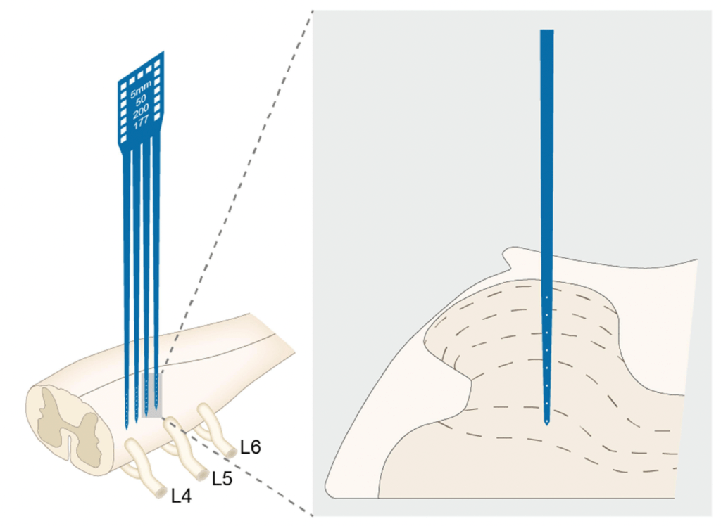
This paper comes from Professor Jing’s team in Beijing and utilises in vivo measurements from the WDR neurones in the SDH of male Sprague-Dawley rats using a rather sophisticated looking microelectrode array.
It attracted me because it tests different locations and intensities of EA and directly measures the activity of related spinal WDR neurones as the outcome.
WDR neurones are essentially the amplification system for nociceptive inputs in the spinal cord. For a patient with an arthritic joint, they can make the difference between a nociceptive input from the joint being stopped at the spinal cord or continuing to the brain and resulting in the perception of pain. It is the sensitivity of these neurones that principally determine the level of pain perception and it is the inhibition of their membranes following acupuncture that can result in pain reduction for weeks.
This paper used both non-noxious and noxious mechanical stimulation of gastrocnemius muscle as an input to find WDR neurones, but then used injection of hypertonic saline (HS) as a noxious test stimulus to measure the frequency response of WDR neurones with a local RF at the site of stimulus. Having identified a stable response from a number of spinal WDR neurones, the team then applied pre-EA at different strengths and locations to see how this would alter the frequency response of the same WDR neurones.
Pre-EA simply refers to EA performed immediately before the test stimulus with HS.
Pre-EA was applied within the local RF of the test site (ie gastrocnemius), in an adjacent non-RF (tibialis anterior), and in a contralateral non-RF (gastrocnemius in the opposite leg).
In each of these sites the pre-EA was applied for 1 minute at 4 different intensities based on the activating thresholds for Aβ, Aδ, and C fibre components of the WDR discharges. The strongest (4th) level of intensity was set at double that for the C fibre component. Starting from 0.1mA at a pulse width of 500μs the thresholds for these different components were set at 0.5mA, 1mA, 2mA, and 4mA for TAβ, ΤAδ, TC, and 2TC respectively.
Injection of hypertonic saline resulted in the frequency of discharge of the WDR neurones jumping from 0.1Hz to ~30Hz. This was reduced by 60% to 70% by pre-EA at ΤAδ, TC, and 2TC intensities, but not by pre-EA at the lower TAβ intensity.
Interestingly, all intensities of pre-EA ‘worked’ when applied to an adjacent non-RF, although the strongest intensity (2Tc) seemed to have the biggest effect.
When it came to the opposite leg (contralateral non-RF), only pre-EA at TC and 2TC intensities had a significant effect of WDR discharges.
This is really useful research that gives us solid physiological support for what we do in clinical practice. It also suggests a level of subtlety that I had not previously considered. Of course, we need to be cautious in assuming that the connections and temporary effects of very brief pre-EA can reflect the longer-term effects we see in clinical practice.
One clinical trial that sits nicely alongside this laboratory research was highlighted on the blog in 2019 – Strong EA and CPM in OAK.[2]
References
1 Yu Q, Cao W, Wang X, et al. The Effect of Pre-Electroacupuncture on Nociceptive Discharges of Spinal Wide Dynamic Range Neurons in Rat. J Pain Res 2023;16:695–706. doi:10.2147/JPR.S396481
2 Lv Z, Shen L, Zhu B, et al. Effects of intensity of electroacupuncture on chronic pain in patients with knee osteoarthritis: a randomized controlled trial. Arthritis Res Ther 2019;21:120. doi:10.1186/s13075-019-1899-6

One thought on “Pre-EA on spinal WDRs”
Comments are closed.