Stimulated by Weagle et al 2020.[1]
POCUS – point of care ultrasound
key to acronyms
POTUS – president of the United States
US – ultrasound
TV – television (the original ones were black and white)
Not another case report of pneumothorax I hear you cry, that is not news to the acupuncture community. It happens from time to time, but it is very rare, what could be new?
Well, I was drawn into this otherwise rather unremarkable report by the mention of POCUS. This was an acronym I had not come across previously, but it was rather like one I had heard recently with a T rather than a C.
POTUS – president of the United States
I heard this term being used frequently in the book A Promised Land; a very interesting account of Barack Obama’s first term as POTUS. I can thoroughly recommend this book, and whilst it is unlikely to improve your acupuncture skills, there is quite a bit of needling going on between the democrats and the republicans.
POCUS – point of care ultrasound
This case report is from Halifax in Canada and the authors are from an emergency medicine department. The full text is available so you can follow this link to read the case and view the linked videos. A 38-year-old woman presented with pleuritic chest pain the day after a session of acupuncture dry needling for back pain. An ECG was unremarkable, and POCUS was used to diagnose a pneumothorax.
I watched the videos that apparently demonstrated the presence of a pneumothorax, but I was unsure what exactly I should be looking at, and the case report mentioned absent lung sliding and the presence of a lung point. Well, that was definitely new to me, so I followed the reference from 2005 to get an idea of the terminology.[2]
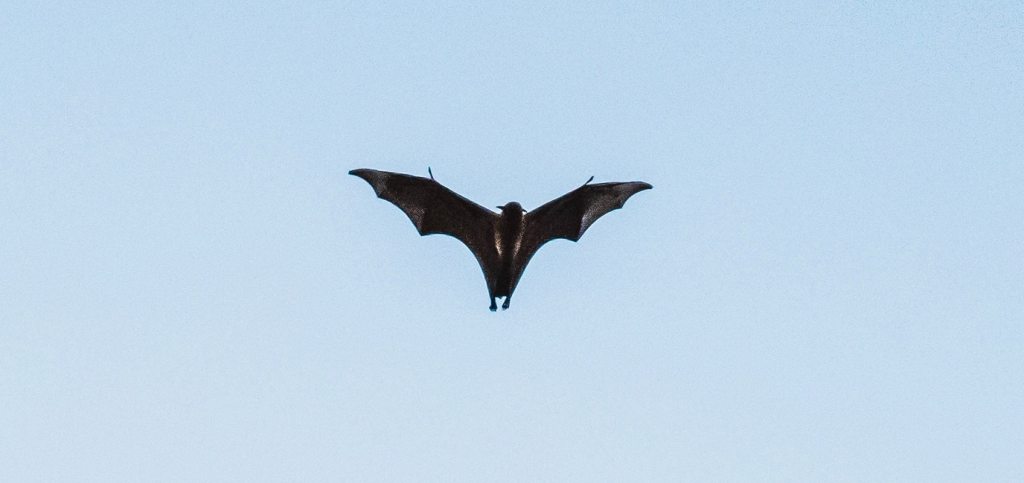
I remember watching batman in black and white on our first TV set when I was in low single figures, probably in the late 60s. So, looking at US images for the ‘bat sign’ was rather similar. US can look rather like a poorly tuned TV, which is the last thing you want to see in the middle of the action. I’m obviously mildly traumatised by the frustration of missing the batman and robin action.
the bat sign
As well as the bat sign, which is the appearance of the pleural line in between the ribs, there are a number of other lines to be identified as well as the crucial lung point.
‘A’ lines are horizontal on the US image, just like the pleural line, but finer and deeper. They represent normal lung tissue. ‘B’ lines are vertical and come in various forms and are associated with some cool names such as comet tail artefacts and lung rockets.
In the normal lung, without air in the pleural space, the expansion of lung during inspiration gives the appearance of movement of tissue below the pleural line. This is referred to as lung sliding and is absent in pneumothorax.
What is the ‘lung point’?
The lung point is the appearance of the point at which the parietal and visceral pleura divide in a pneumothorax, which means that the lung tissue is no longer in contact with the chest wall and the appearance below the pleural line abruptly changes from the normal granular appearance to a pattern of horizontal lines.
In this case the pneumothorax became symptomatic between about 12 and 24 hours after dry needling the back. It appeared on the subsequent chest x ray film to be relatively small and apical – small pneumothoraces are generally apical of course. I was slightly surprised to read that the team inserted a chest drain to treat this rather small pneumothorax, particularly as the patient had no shortness of breath.
I have seen a rather similar sized apical pneumothorax diagnosed the day after a demonstration of dry needling on the back, and in this case no treatment was required. The air in the pleural space was absorbed spontaneously, although I am unsure how long that took to happen as we did not perform regular imaging, and the routine follow up was delayed.[3]
References
1 Weagle K, Henneberry RJ, Atkinson P. Pneumothorax Following Acupuncture. Cureus 2021;13:e14207. doi:10.7759/cureus.14207
2 Lichtenstein DA, Mezière G, Lascols N, et al. Ultrasound diagnosis of occult pneumothorax. Crit Care Med 2005;33:1231–8. doi:10.1097/01.ccm.0000164542.86954.b4
3 Cummings M, Ross-Marrs R, Gerwin R. Pneumothorax complication of deep dry needling demonstration. Acupunct Med 2014;32:517–9. doi:10.1136/acupmed-2014-010659

You must be logged in to post a comment.